Cartilage defects of the Knee Joint
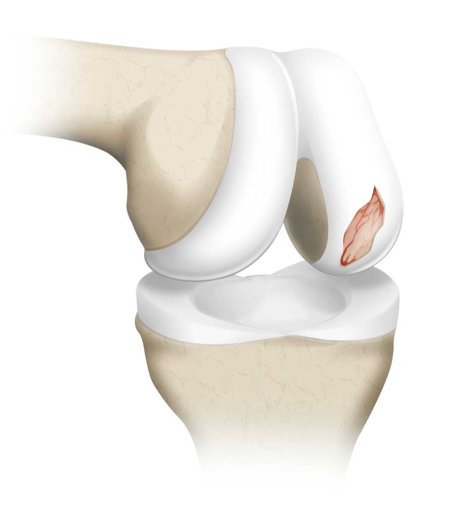
Cartilage defects in the knees are damage to the articular cartilage, the smooth material that covers the ends of the bones to prevent them from rubbing against one another. Degenerative cartilage defects are the consequence of wear and tear, while traumatic cartilage defects are brought on by an injury such as a fall onto the knee, a jump, or a sudden change in direction while playing a sport. Due to the lack of nerves in cartilage, such injuries do not necessarily result in acute symptoms. However, over time, cartilage issues can impair normal joint performance, resulting in discomfort, inflammation, a grinding sensation in the knee, and restricted mobility.

Symptoms of Cartilage Defects.

Individuals who have joint cartilage damage will suffer from:
- An area experiencing inflammation will enlarge, become warmer than other parts of the body, and feel uncomfortable, tender, and sore.
- Stiffness in the knee joint.
- Range restriction: when the injury worsens, the afflicted limb will become less mobile.
The knee is the joint where articular cartilage injury happens most frequently, but it can also affect the elbow, wrist, ankle, shoulder, and hip joints.
How are knee cartilage defects diagnosed?
-
Questions asked by Doctor
The first questions the doctor will ask you relate to pain and other symptoms in and around the injured joint. Inquiries regarding prior surgeries, whether a particular injury triggered the symptoms, and the kinds of physical activity usually engaged in may also be made.
-
Physical Examination
The physician will watch the joint’s motion during the physical examination. To assess pain, edema, range of motion, and ligament stability, he or she may rotate and bend the joint. Additionally, he or she could instruct the patient to move in specific ways; for example, if the patient has a knee injury, they might be instructed to walk, squat, or “duck walk.”
-
X-ray/ MRI
It might also be necessary to do imaging tests, such as a magnetic resonance imaging (MRI) examination or a weight-bearing X-ray.
-
Other Diagnosis
An arthroscopy is occasionally performed to aid in the diagnosis of articular cartilage disease or injury. A tiny incision or two is made on the skin surrounding the injured joint during an arthroscopy, a minimally invasive surgical technique in which the surgeon inserts surgical instruments and an arthroscope—a thin, pencil-sized tube with a camera—into the joint. The surgeon can see the tissues and structures of the joint by watching a video feed from the camera that is sent to a monitor.
Treatment of Cartilage Defects
1) Debridement
Debridement may be an option for older patients with minor symptoms and lesser cartilage abnormalities.
2) Microfracture
An arthroscopic treatment called microfracture is used to restore damaged knee cartilage.
3) Osteochondral Autograft Transplantation (OATS)
In this procedure, healthy cartilage is removed from a patient’s non-weight-bearing area and transplanted to the damaged area.
4) Autologous Chondrocyte Implantation
A sample of healthy cartilage is taken out during this process, reproduced in large quantities outside the body, and then re-implanted onto the nearby bone. This recently developed cartilage wraps the bone, providing support and protection.
